.png)
Pet Imaging Centre (PICs)
.png)
6 BROOKS AVE Wyoming 2250. Ph 4329 0500
MENU

Please consider Pet Insurance while your pet is healthy & phone for advice on your options.

Toby didn't want to play ball anymore -
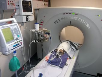
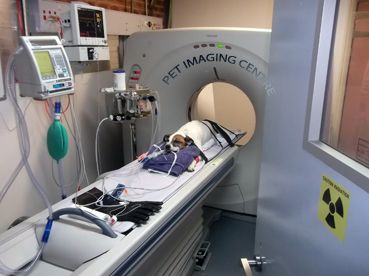
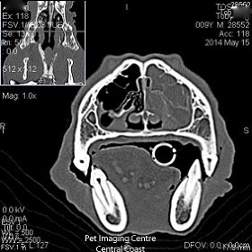
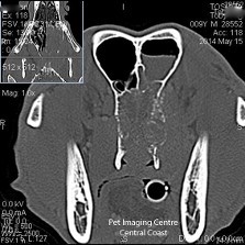
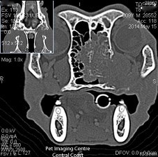
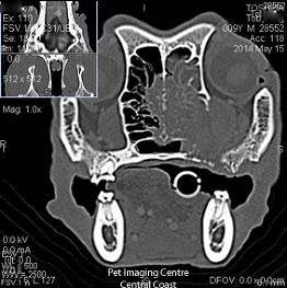
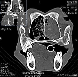
Toby presented to their local vet 5 weeks before the CT scan. He resented his mouth & jaw being opened, he did not like playing with his ball as much & was occasionally reverse sneezing. At one point his eye drooped slightly then appeared normal again. A dental was performed and a few rear upper loose molars were removed. Dental radiographs at the time showed otherwise nice teeth & roots. Toby did not improve after the dental & stopped playing ball. Clinical signs also did not improve with antibiotics so radiographs of the nasal cavity & skull were performed under general anaesthetic. An unusual area was seen on radiographs and a CT scan ordered. PICs performed a contrast CT of the nasal cavity. Based on the history, we extended the fine 1mm scan to include the termporal mandibular joint (jaw joint or TMJ), the middle ear, the brain and the pharynx & larynx. A chest scan was also performed.
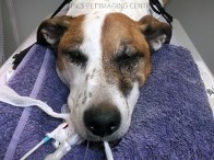
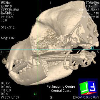
The specialist CT report concluded there is a large soft tissue mass causing severe bone lysis (eating away the bone), obliterating the left nasal cavity. The bone is disappearing from the left & right nasal turbinates, the nasal septum (the midline), the cribriform plate (between the nasal cavity and the brain), the maxillary bone both laterally (under the eye) & ventrally (towards the mouth). Conclusions from the CT scan were likely adenocarcinoma (cancer) originating in the left nasal cavity with extension into the right nasal cavity, left retrobulbar space (under the eye), left maxillary region and through the cribriform plate- extending into the frontal lobe of the brain. With the help from hind-sight, the two molar teeth found loose at the dental were most likely secondary to the adjacent bone lysis from the nasal tumor.Toby was returned to his local vet for treatment options. We remember him for his fighting spirit & affection.
The specialist CT report concluded there is a large soft tissue mass causing severe bone lysis (eating away the bone), obliterating the left nasal cavity. The bone is disappearing from the left & right nasal turbinates, the nasal septum (the midline), the cribriform plate (between the nasal cavity and the brain), the maxillary bone both laterally (under the eye) & ventrally (towards the mouth). Conclusions from the CT scan were likely adenocarcinoma (cancer) originating in the left nasal cavity with extension into the right nasal cavity, left retrobulbar space (under the eye), left maxillary region and through the cribriform plate- extending into the frontal lobe of the brain. With the help from hind-sight, the two molar teeth found loose at the dental were most likely secondary to the adjacent bone lysis from the nasal tumor.Toby was returned to his local vet for treatment options. We remember him for his fighting spirit & affection.
PET IMAGING CENTRE (PICs) / CCVC (02) 43 29 0500PICs CCVC 6 Brooks Avenue, Wyoming, NSW 2250, AUSTRALIA