.png)
Pet Imaging Centre (PICs)
.png)
6 BROOKS AVE Wyoming 2250. Ph 4329 0500
MENU

Please consider Pet Insurance while your pet is healthy & phone for advice on your options.
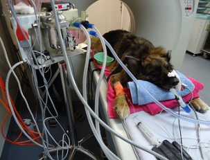
SASHA THE GERMAN SHEPHERD
A routine screening CT reveals an immediate life-threatening condition!
Sasha, a desexed 9 yr old female German Shepherd, presented a month prior. She was struggling on her back legs, stiff and did not want to rise in the morning. A complete blood test was normal and anti-inflammatories were prescribed. She was "better than ever since starting anti-inflammatories", brighter and jumping up into the car. She was still reluctant to eat, but had always been a fussy eater. A month later she represented as not being quite right, not eating alot, panting and fatiguing on the walk. Her heart rate was 130 bpm which is high for her when at rest. The owner agreed to have a CT performed as a general scan, specifically focusing on the chest. A complete blood test again came back normal.
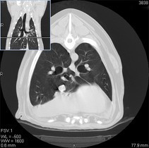
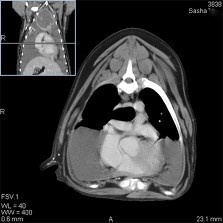
A contrast CT study was performed at PICS. Chest and abdomen, along with the hips were scanned. Immediately following the first chest CT, the scan was interrupted and the owner was phoned. She was advised a cystic mass in the front of the chest (cranial Mediastinum) was found and half the chest was filled with fluid. Permission was granted to drain both sides of the chest and over 1litre of liquid was removed.
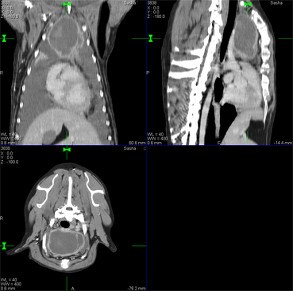
A repeat CT scan of the chest was performed free of charge by the vets, immediately after draining. This was in order to have the best images for specialist review. The fluid drained was sent away for analysis.
The CT specialist reported that the cystic mediastinal mass was placing pressure on the vascular/nervous bundles in the chest, with concurrent bilateral pleural effusion (fluid within the chest). Other spinal conditions like spondylosis were noted along with lymph node involvement and a list of the most likely causes of the mass. The fluid analysis from the chest returned no cancer cells.
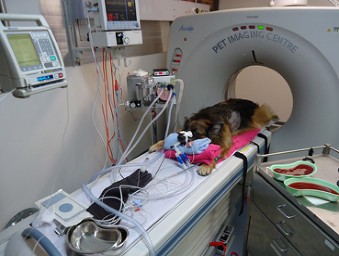
More than 1 Litre of BLOOD TINGED FLUID was drained from both sides of the chest (see Kidney dishes at top).
Next was to biopsy the mass itself for pathology. An ultrasound guided Fine Needle Aspirate was performed, actually taking a sample of a smaller tissue mass within the front cyst. The pathology of the tissue returned as necrotic or dead tissue (so we still did not have the exact cause). Open chest surgery & complete surgical removal was successfully performed. The mass and associated mediastinal lymph node was submitted for histopathology. This revealed findings consistent with a thymoma (predominantly epithelial) and reactive lymph node hyperplasia, which was great news rather than a cancer. With it removed a full recovery was expected. An extended period of chest draining was required for up to 4-6 wks post surgery, after which Sasha did make a full recovery and she should lead a normal healthy life
PET IMAGING CENTRE (PICs) / CCVC (02) 43 29 0500PICs CCVC 6 Brooks Avenue, Wyoming, NSW 2250, AUSTRALIA

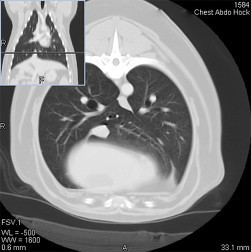
The images above & below show the difference post-draining. The LUNGS can now be seen expanding down lower and on each side of the heart. Throughout, the anaesthetic the oxygen blood levels remained good.