.png)
Pet Imaging Centre (PICs)
.png)
6 BROOKS AVE Wyoming 2250. Ph 4329 0500
MENU

Please consider Pet Insurance while your pet is healthy & phone for advice on your options.
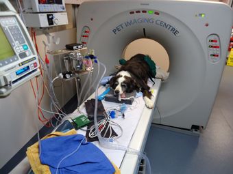
Sam the Border Collie -
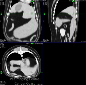
Not quite right, lethargic, vomiting and an old stiff joint- it was time to perform a General Health CT Screen to work out his list of issues.
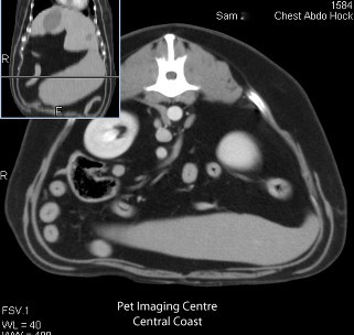
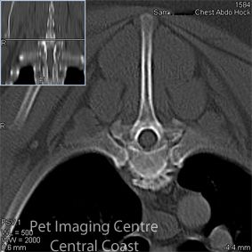
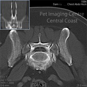
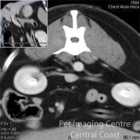
We have known Sam since 2006. Over time we helped him through being sick from a big cooked Xmas-Veal Bone back in '07, bad joints from taking on a truck, running back crying from the bushes in the park and other odd lumps & bumps along the way. Now he was vomiting on and off for a few days & not improving. We knew his right hock was stiff and he had a severe decrease in range of motion from the past. Blood testing a month prior had shown his liver levels were mildly increased but had resolved at retesting a few weeks later. However, he still had some circulating nucleated red blood cells which in the absence of anaemia was unusual. The spleen needed to be checked & what was the cause of the vomiting? So the decision was to skip past x-raying all these areas- back legs, chest, abdomen & spine, & just perform a general CT, costing about around the same & taking a lot less time under anaesthetic, as well as producing much better images. He is also at the age where such a CT health check could be very beneficial regardless. His COMPLETE SPECIALIST CT REPORT is shown below, to give you an idea of how thorough a General Health CT Scan can be while they are under a short, heavy sedation or anaesthetic. For Sam, we injected contrast to have very clear pictures or his arteries, veins and kidneys. In Sam's cases it revealed a mild enlargement of his left atrium of his heart, diffuse mural gastropathy (stomach problem), focal hepatic lesion (lesion in his liver -see top left picture) and severe bilateral Degenerative Joint Disease (DJD) of both hocks. Severe spondylosis along a number of his vertebra and a disc protrusion in to his spine at the level of L7-Sacrum. So this helped the vets immensely. The severe gastritis was controlled with multiple targeted medications, the very mild flucuating liver enzymes could be from the nodular liver lobe changes & needs ongoing monitoring. In addition, we learn't about his heart, his spine and his hock condition. As seen below, it also checked and ruled out many problems, it was great that we did not find any major concerns, although his stomach wall was not biopsied in the end. Sam did return to his normal self after some medication & did not require a gastroscope to check or biopsy his stomach. His heart needs to be monitored for changes in chamber sizes & his heart values evaulated with echocardiography in our ultrasound department. This report is a great reference for the future for the owner, the vets involved and allows targeted treatment fo each of his many conditions.
CT Imaging Study Report PICs Radiologist Exam Date ##-##-2013 Report Date ##-##-2013 Patient number ##### Patient Name Sam ##### 10 yr oldMN Border collie History ·Previous vomiting bouts ·Not quite right- vague, not eating as much ·Persistant occasional nucleated RBC on the bloods ·Injury to the R hock- thickening decreased ROM Modality& Exam Post contrast CT of the thorax and abdomen. CT of the tarsi Findings Thorax Mild enlargement of the left atrium. Normal lung vessels and overall density No evidence of mediastinal, thoracic wall or pleural abnormalities Abdomen The stomach is only minimally distended with fluid content. Generalised thickening of the gastric wall. Rugal folds cannot be identified. Focal hypodense image located in the left medial liver lobe. This structure measures around 2 cm in diameter. Unremarkable gallbladder and common bile duct Pancreas with normal shape, density, contours and size Small rounded soft tissue dense structure located immediately ventral and lateral to the descending duodenum The jejunum and colon present normal overall diameter and content. Normal adrenal glands, kidneys and urinary bladder No evidence of free abdominal fluid Severe spondylosis and disc protrusion L7-S Right tarsus Severe perarticular new formation involving the talocrural and proximal inter-tarsal joints. No evidence of lysis in the subchondral or metaphyseal bone Left tarsus Similar, but less severe, changes compared to the right tarsus Summary: Diffuse mural gastropathy, focal hepatic lesion and server bilateral DJD tarsi Interpretation: Findings in the stomach are most likely consistent with severe gastritis. Differential Diagnosis (DD) could include diffuse neoplastic infiltrate (lymphosarcoma) Findings in the liver are most likely consistent with nodular regeneration. Less likely DD include metastasis, cyst, abscess Recommendations Gastroscopy and gastric wall biopsy. Cardiac echocardiography Biopsy or monitor liver levels.

.jpg)
PET IMAGING CENTRE (PICs) / CCVC (02) 43 29 0500PICs CCVC 6 Brooks Avenue, Wyoming, NSW 2250, AUSTRALIA